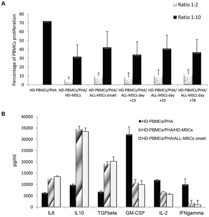
Figure 5. A: In vitro immunomodulatory effect of HD-MSCs and ALL-MSCs on HD-PBMCs.
The graph shows the proliferation of healthy donor peripheral blood mononuclear cells (HD-PBMCs) stimulated with phytohemagglutinin (PHA) either in the presence or in the absence of HD-MSCs or ALL-MSCs, generated at disease onset and subsequent time-points of treatment (day+15; day+33; day+78). Each bar represents the percentage of proliferation of 105 PBMCs, in the presence of two different MSC∶PBMC ratios (MSC∶PBMC ratio of 1∶2 and 1∶10), calculated by measuring 3H-thymidine incorporation after 3-day co-culture. The counts per minute (cpm) values at each cell concentration were normalized to the cpm of PBMCs without MSCs in each experiment. Each bar represents the mean ± SD of multiple experiments (each point being in triplicate) with MSCs obtained from 10 ALL patients (at each time-point) and 10 HDs. B: Quantification of cytokines and growth factors in supernatants of co-cultures of HD-MSCs and ALL-MSCs (isolated at day+0) with PBMCs, after 72-hour incubation with PHA. An increase in anti-inflammatory cytokines (i.e. IL6, IL10 and TGFb) was detected in the presence of both HD-MSCs and ALL-MSCs, as compared with PHA-stimulated PBMC cultures. A decrease in pro-inflammatory cytokines and growth factors (GM-CSF, IL2 and IFNg) was revealed in supernatants collected from the same co-cultures. Each bar represents the mean +/−SD of the results from experiments performed with the same HD- and ALL-MSC samples employed in the PBMC proliferation assays. Results are expressed as pg/ml.

