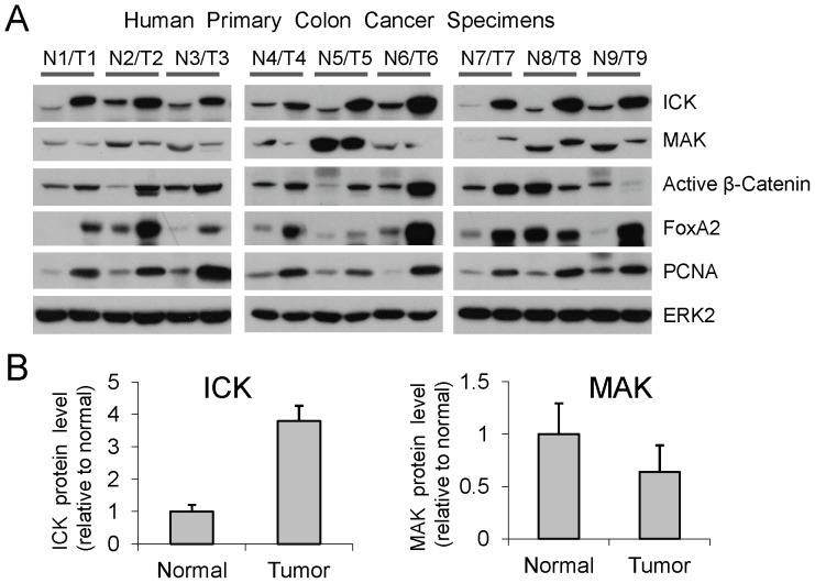
Figure 6. ICK and MAK proteins exhibited distinct expression patterns in human colorectal tumors.
(A) Tissue extracts from nine pairs of human specimens macro-dissected from primary colon tumor (T) and its surrounding normal mucosa (N) were prepared. Equal amount of total proteins (30 µg) from tissue extracts were loaded for Western blot. ERK2 signals indicate equal loading of total proteins. PCNA was used as a proliferation marker. (B) Western blot signals of ICK and MAK were quantified by densitometry and normalized against ERK2 signals. ICK and MAK protein levels in tumors relative to their adjacent normal tissues were shown as mean ± SE, n = 9, P<0.05, Paired t-Test.

