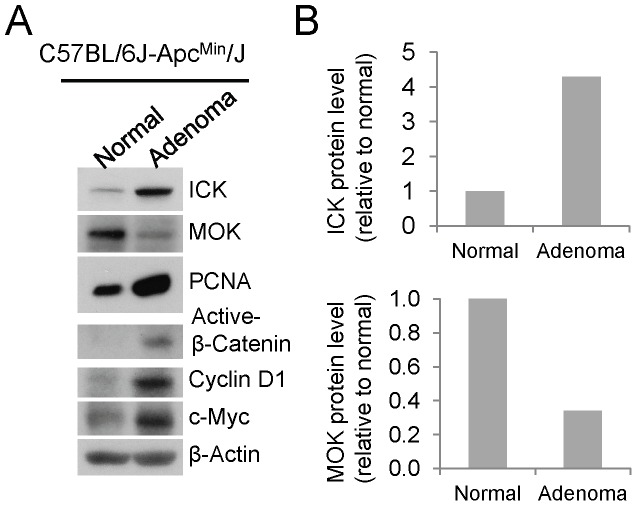
Figure 7. ICK and MOK proteins displayed opposite expression patterns in mouse intestinal adenomas.
(A) Tissue specimens macro-dissected from intestinal adenomatous polyps and their adjacent normal mucosa were obtained from C57BL/6J-Apcmin/J mice (The Jackson Lab). Equal amount of total proteins (30 µg) from tissue extracts were Western blotted against ICK and MOK antibodies respectively. PCNA signal was shown as a proliferation marker to indicate malignant proliferation in the intestinal adenomas. Western signal of β-Actin was shown to indicate equal loading of total proteins. (B) Western blot signals of ICK and MOK were quantified by densitometry and normalized against the β-Actin signal. Shown here were the protein levels of ICK and MAK in intestinal adenomas relative to their adjacent normal tissues.

