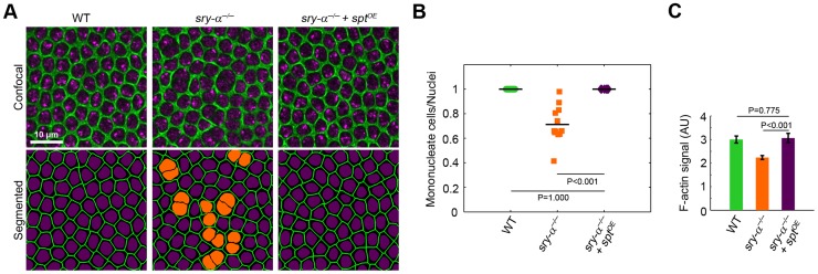
Figure 4. Sry-α and Spt share redundant function during cellularization.
(A) Surface views of furrow canals stained for Myosin-2 in embryos of indicated genotypes. Multinucleate cells highlighted (orange nuclei) in corresponding segmented images. (B) Quantification of multinucleation phenotypes. Each point represents one embryo with ≥150 nuclei analyzed (n = 12 embryos per condition). (C) Quantification of F-actin in furrow canals of furrows of length 3–5.5 µm (n = 9 embryos per genotype, with 15 furrows analyzed per embryo; mean ± s.e.m.). Student's t-test was performed to calculate P values as shown in (B, C).

