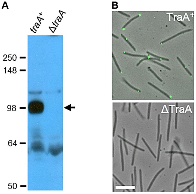
Figure 3. TraA is a cell surface receptor.

A) Western blot with TraA-PA14 antibodies against whole-cell lysates from traA + (DW1463) and ΔtraA (DW1467) strains. Molecular weight markers (kDa) are shown at the left, and the arrow indicates the TraA-specific band at ∼100 kDa. B) TraA immunofluorescence micrographs of live non-permeabilized cells. The same strains and primary antibodies were used as in A. White bar represents 2 µm.
