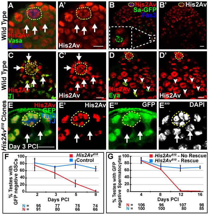
Figure 1. His2Av is required cell autonomously for GSC maintenance.
(A–A′): Apical tip of wild-type testis immunostained with anti-His2Av (red), anti-Vasa (green), and anti-Fas3 (blue). GSCs (arrows) identified as Vasa positive cells adjacent to the hub (yellow dashed line). (B–B′): Testes expressing Sa-GFP (green) to mark spermatocytes, immunostained with anti-His2Av (red), anti-GFP (green), and anti-Fas3 (blue). Inset shows spermatocyte nucleus with Sa-GFP marking the nucleolus and His2Av localized to the chromosomes; hub (yellow dashed line). (C–D′): Apical tip of wild-type testes immunostained with anti-His2Av (red, C–D′), anti-Tj (green, C) to mark CySCs (arrows, C, C′) adjacent to the hub (yellow dashed line) or anti-Eya (green, D) to mark cyst cells (arrowheads, D, D′). (E–E′″): Testes day 3 PCI immunostained with anti-GFP (green) to identify homozygous His2Av810 mutant GSCs (arrows), anti-His2Av (red), anti- Fas3 and Tj (blue) and DAPI (E′″); hub (yellow dashed line). Scale bars: 10 µm (F): Percentage of testes with His2Av810 mutant (red) or FRT 82B control (blue) GSCs scored at indicated times PCI.(G): Percentage of testes with His2Av810 mutant spermatocyte cysts in a genetic background either with (blue line, rescue) or lacking (red line, no rescue) a His2Av-mRFP genomic rescue transgene. Data shows average ± S.D.

