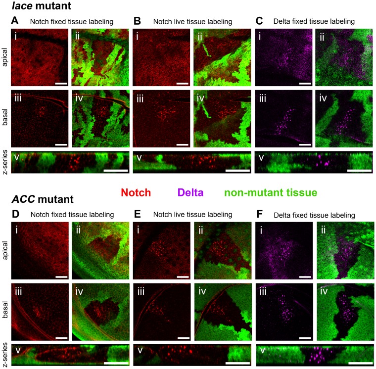
Figure 1. Notch and Delta accumulate abnormally in lace and ACC mutant tissues.
Confocal optical sections through D. melanogaster wing imaginal discs bearing homozygous mutant clones of lace18 (A–C) or ACC1 (D–F) showing accumulation of Notch (A, D) and Delta (C, F) in fixed tissue samples, and endocytic internalization of Notch in live tissue samples (B, E). Areas devoid of GFP marker gene expression (green) correspond to mutant cell regions. Each set of five images (i–v) depict an apical (i, ii) and basal (iii–iv) horizontal section showing Notch (red in A, B, D and E) or Delta (magenta in C and F) accumulation, the same images overlaid with corresponding GFP expression to indicate clone locations (ii and iv), and a representative z-series showing the distribution of Notch or Delta along the apicobasal axis of the disc tissue (v). Scale bars, 20 µm.

