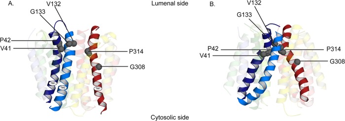
FIGURE 9.

Clustering of the mutations in the lumenal (A) and cytoplasmic (B) facing model of rVMAT2. Helices are shown as schematics and viewed along the plane of the membrane with the cytoplasm to the bottom. The key helices, TM1, -2, and -7, are opaque and all the others are transparent.
