
FIGURE 1.
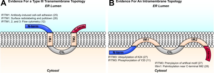
Two models of IFITM protein topology are shown. Evidence for the orientation of each terminus with respect to the membrane is shown with the relevant citation. A, IFITM proteins were predicted to assume a two-pass type III transmembrane topology, with two transmembrane domains (TM1 and TM2) flanking the conserved intracellular loop (CIL). Early studies identified IFITM1 N termini in the extracellular space, and other investigators have detected IFITM N termini at the plasma membrane by flow cytometry. (See figure for references.) B, more recent studies support a topology with two intramembrane domains (IM1 and IM2) and cytosolic N and C termini. Post-translational modifications of the IFITM3 N terminus imply access of the N terminus to cytosolic enzymes. Evidence for an intracellular C terminus comes from studies of lipidation motif reporters. (See figure for references.)