FIGURE 2.
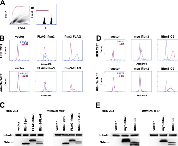
FLAG-tagged Ifitm3 N and C termini can be detected at the surface of intact cells. A, the gating strategy used to produce subsequent histograms is shown here. To confirm staining of only extracellular FLAG antigen, PI-positive cells were excluded from analysis. SSC-A, side scatter; FSC-A, forward scatter. B, staining for the FLAG epitopes on 293T cells and MEFs is shown in blue. Fluorescence staining from the isotype control is shown in red. Alexa488, Alexa Fluor 488. C, expression levels of wild type and N- and C-terminally FLAG-tagged Ifitm3 were determined by Western blot with an antibody against the native Ifitm3 N terminus. D, the experiment shown in A was repeated with N-terminal Myc and C-terminal C9 tags in place of the FLAG epitopes. Myc staining is shown in blue, and C9 staining is shown in red. E, total expression of the constructs was determined by Western blot. The double band pattern for C-terminally tagged Ifitm3 constructs is explored in detail in Fig. 6.

