FIGURE 3.
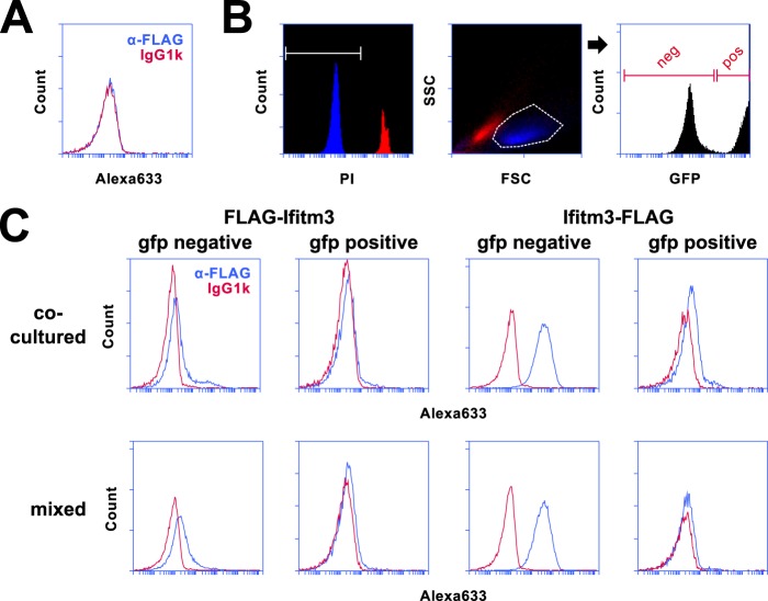
Surface staining of N and C termini is not an artifact of membrane adherent cellular debris. A, staining of Ifitm3−/eGFP+ with anti-FLAG (blue) and isotype control (red) antibodies is shown. Alexa633, Alexa Fluor 633. B, PI− (intact) and PI+ (permeabilized) 293T cell populations were mapped onto a forward/side scatter plot (FSC/SSC). Intact 293T cells (blue) could be clearly distinguished from cells with compromised membrane integrity (red) by scatter gating alone. Scatter gating was used in lieu of PI staining to ensure analysis of only intact cells for during subsequent experiments. Cells were further sorted based on eGFP expression as shown in the rightmost histogram. C, cells expressing either FLAG-Ifitm3 or Ifitm3-FLAG were co-cultured with Ifitm3−/eGFP+ cells for 18 h (top panels) or mixed with Ifitm3−/eGFP+ cells immediately prior to surface staining (bottom panels). FLAG staining is shown in blue, and isotype control staining is shown in red.

