FIGURE 9.
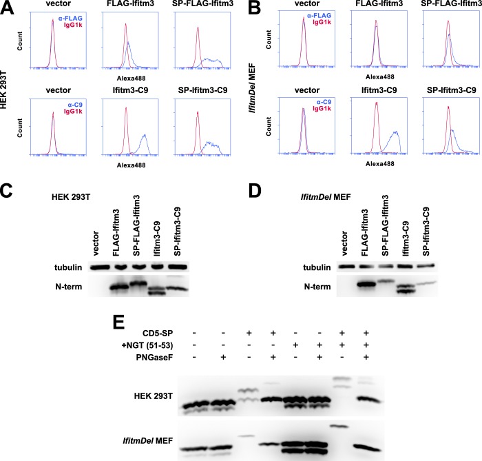
The addition of a signal peptide to the Ifitm3 N terminus mimics a type III topology. A, 293T cells expressing FLAG-Ifitm3, Ifitm3-C9, or the same constructs with the addition of an N-terminal CD5 signal peptide were stained for the appropriate epitopes and analyzed by flow cytometry. Alexa488, Alexa Fluor 488. B, MEFs were analyzed by flow cytometry as above. C, a Western blot for the native Ifitm3 N terminus shows the relative expression of each construct in 293T cells. Lysates were treated with peptide-N-glycosidase F to remove N-linked glycosylation of the N terminus. D, the relative expression of each construct in MEFs is shown by Western blot. E, the N terminus of Ifitm3 can be glycosylated but is not normally accessible to cellular glycosylation machinery. Cells were transfected (293T, top) or transduced (MEF, bottom) with constructs expressing C9-tagged Ifitm3 with or without a CD5 signal peptide (CD5-SP) and with or without introduction of an additional NGT motif (+NGT (51–53)). Lysates were treated with peptide-N-glycosidase F (PNGaseF) or mock-treated as indicated and analyzed by Western blot with antibodies against the native N terminus.

