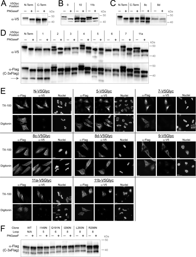
FIGURE 6.
V5Glyc tag allows mapping of GOAT topology using two different techniques. A–D, glycosylation gel shift assay for localization in transfected HeLa cells. Mouse GOAT constructs with internal or terminal V5Glyc tags and a constant C-terminal 3xFLAG tag were lysed 20 h after transfection and treated with peptide-N-glycosidase F (PNGaseF) or mock-treated in identical buffer and then subjected to anti-V5 immunoblotting. Only lumenal positions can be glycosylated, and peptide-N-glycosidase F treatment cleaves off the oligosaccharide. Constructs in B were less well expressed than those in D. The anti-FLAG blot in D shows an additional band present (gray arrow) not seen in the anti-V5 blot for the N-terminal construct and position 1; this band is shown below to represent a protein starting with Met-56. Note that positions 8a and 8b are not shown; no full-length GOAT could be detected from these constructs. In C, two additional positions were tested in loop 8 (8c and 8d), and both expressed well and confirmed cytosolic location. E, selective permeabilization of key V5Glyc constructs reports lumenal location for challenging positions 9 and 11 and cytosolic location of loop 8; other previously suggested positions are also confirmed. As above, GOAT bearing the indicated internal V5Glyc tag was used, and all constructs have a constant C-terminal 3xFLAG tag. The N terminus and positions 5 and 7 are shown because these were the most robust in our initial survey. F, NXS or NXT codons (not V5Glyc tags) were installed in all loops longer than 24 amino acids: loops 5, 6, and 8. Loop 8 is longer, so three constructs were generated. None of these epitopes could be glycosylated, consistent with cytosolic locations.

