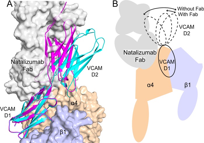
FIGURE 6.
Model of VCAM binding to a natalizumab Fab-α4β1 complex. Transparent solvent accessible surfaces are shown of the α4 β-propeller domain (wheat), β1 βI domain (light blue), and natalizumab Fab (gray). Two examples of VCAM D1D2 crystal structures (chain A from Ref. 28 and chain B from Ref. 29) that differ the most in D1-D2 orientation are shown in ribbon representation, docked as described (11). The model of α4β1 is described in the legend to Fig. 5. Strong clashes show regions where the schematic is largely obscured by the transparent surface.

