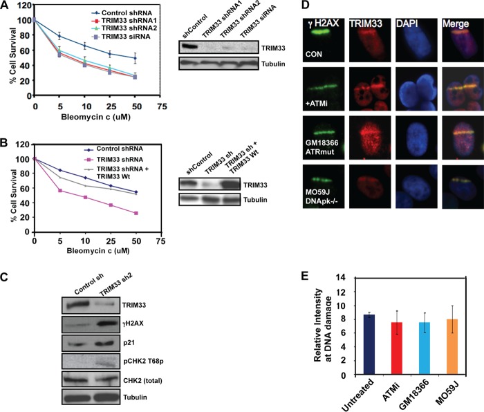
FIGURE 3.
TRIM33 knockdown results in DNA damage sensitivity. A, HeLa cells treated with control shRNA, TRIM33 shRNA1, TRIM33 shRNA2, and TRIM33 siRNA were exposed to increasing concentrations of bleomycin. Relative cell counts measured by MTS assay, normalized to no treatment, were performed on day 3 and were plotted. p < 0.005 for control versus TRIM33 shRNA or siRNA. Relative expression of TRIM33 and tubulin is shown. B, HeLa cells treated with control shRNA, TRIM33 shRNA, or TRIM33 shRNA cells complemented with WtTRIM33 were exposed to increasing concentrations of bleomycin, and relative cell counts were measured as above. The Western blot analysis shows levels of TRIM33 and tubulin. C, whole cell extracts from control and TRIM33 sh2-treated cells were processed for Western blotting using antibodies to the indicated proteins. D, TRIM33 localization to DNA damage is not dependent on ATM, ATR, or DNA-dependent protein kinase (DNApk). HeLa cells treated with vehicle or ATM inhibitor (ATMi) (KU-55933), GM18366 (ATR mutant), and M059J (DNApk−/−) cells were subject to laser scissor-induced DNA damage. Cells were fixed after 10 min and processed for IF using antibodies to γH2AX (green) and TRIM33 (red). E, quantitation of TRIM33 at sites of DNA damage is shown. Each data point is the mean ± S.D. of at least 20 cells.

