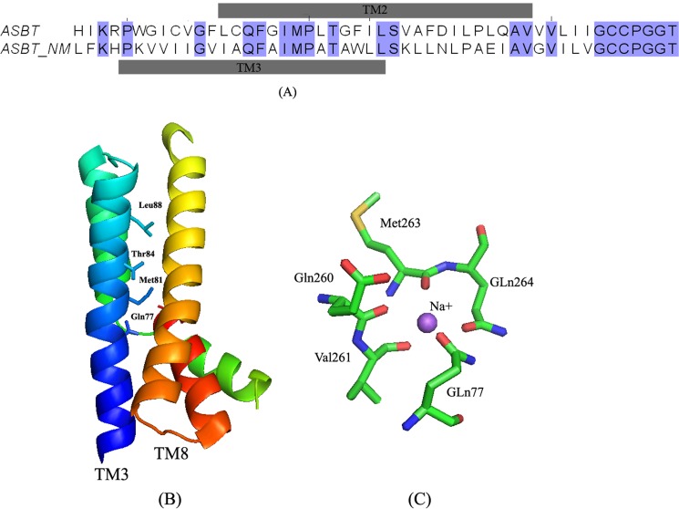
FIGURE 10.
Sequence alignment and mapping of TM2 sodium sensitive residues onto the ASBTNM structure. A, sequence alignment of TM2 and its flanking residues of hASBT to ASBTNM using ClustalW. The alignment results were manually adjusted in JalView 2.8. The gray bars above and below the sequence pair indicate the amino acid range of each transmembrane domains. Residues that are identical in both sequences are highlighted in blue. B, ribbon representation of TM3 and TM8 structures of ASBTNM with residues shown to correspond to the sodium-sensitive residues in TM2 of hASBT. C, stick model of the amino acids that mediate sodium binding in the crystal structure. Amino acids are colored by elements: green for carbon, blue for nitrogen, and red for oxygen.

