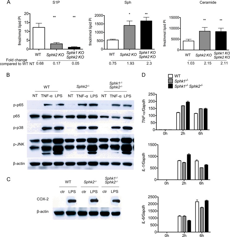
FIGURE 3.
Sphk isoenzymes are not necessary for inflammatory responses. A, intracellular sphingoid bases and total ceramides in TNFα-treated BMDMs quantified by LC-MS/MS. *, p < 0.05; **, p < 0.01 (compared with the WT group). n = 4 per group. Data represent mean ± S.E. (error bars). B, immunodetection of total p65 and phosphorylated forms of p65, p38, and JNK in whole cell extracts of BMDMs. Cells were either stimulated or not with 10 ng/ml TNFα for 15 min or 100 ng/ml LPS for 30 min. C, immunodetection of COX-2 in whole cell extracts of BMDMs stimulated or not with 100 ng/ml LPS for 24 h. D, qRT-PCR detection of TNFα, IL-1, and IL-6 mRNA in BMDMs cultured with 100 ng/ml LPS for 0, 2, and 6 h. Bar graphs present data as -fold changes relative to untreated (0 h) cultures (n = 9). Data represent mean ± S.E. (error bars).

