FIGURE 3.
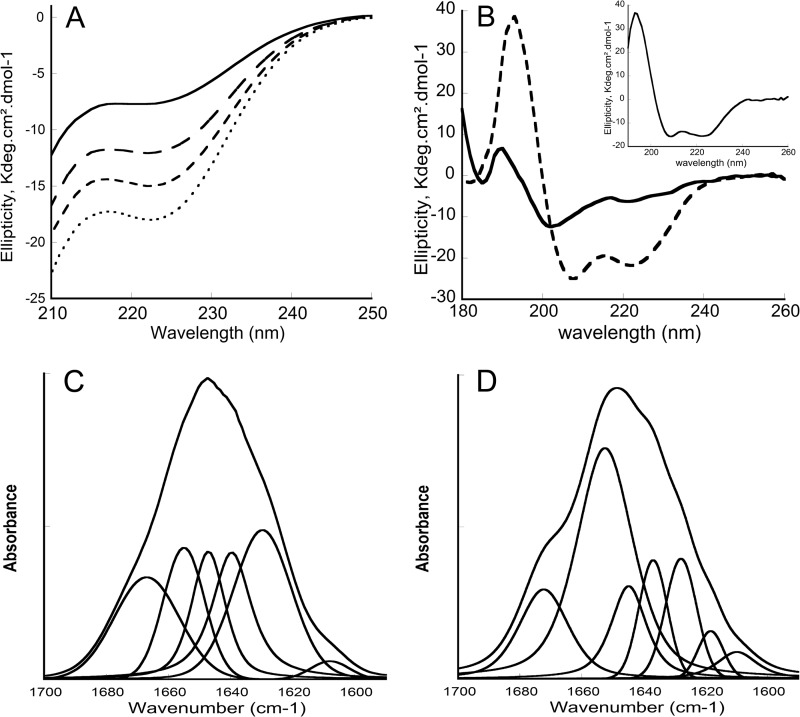
Circular dichroism and FTIR spectra of P454. A, far-UV CD spectra of P454 (40 μm) in the absence (solid line) or in the presence (dashed lines) of increasing concentrations (0.5, 1, and 2 mm from top to bottom) of anionic SUVs made of DOPC/DOPE/DOPG/cholesterol (4:3:2:1) in buffer B at 25 °C. B, synchrotron radiation CD spectra of P454 (200 μm) in the absence (solid line) or in the presence (dashed line) of anionic SUVs (10 mm) in buffer B at 25 °C. C and D, transmission FTIR spectra and band deconvolutions of P454 (150 μm) in D2O buffer B (C) or in the presence (D) of anionic SUVs (5 mm lipids) in D2O buffer B at 25 °C. The S.D. value is within 5%. Secondary structure content analysis is reported in supplemental Table S3.

