FIGURE 8.
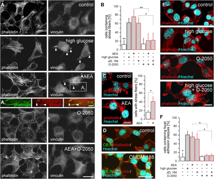
CB1R activation induces cytoskeletal remodeling. A, exposure to glucose (15 mm) or AEA (10 μm, 10 min) induced stress fiber and FA plaque formation in INS-1E cells. O-2050 reversed AEA-induced cytoskeletal modifications. A1, FA plaques localized at the plus ends of polymerized F-actin, providing cytoskeletal anchor points. B, quantitative assessment of the percentage of INS-1E cells with stress fibers upon treatment with AEA (30 min) or JZL 184 (24 h) stimuli alone or in combination with O-2050 (100 nm as 10 min pre-treatment). C, INS-1E cells were exposed to AEA (10 μm) for 24 h to verify that the induction of insulin release by both endocannabinoids upon prolonged activation (see also Fig. 5, A and B) involves stress fiber formation. D, OMDM 188, a DAGL inhibitor (24 h), increased CB1R immunoreactivity in INS-1E cells. E, high glucose (15 mm for 10 min) induced stress fibers in INS-1E cells, as revealed by staining for F-actin (phalloidin). This effect was not reversed by O-2050, a CB1R antagonist. F, quantitative assessment of the percentage of INS-1E cells with FA plaques from experiments as above. High glucose (15 mm) was used as positive control. Data were expressed as mean ± S.D.; n = 30–80 cells/condition were analyzed in triplicate experiments. **, p < 0.01; *, p < 0.05 (pairwise comparisons following one-way ANOVA). Scale bars = 20 μm (F), 10 μm (A, C, and E), and 3 μm (A1).

