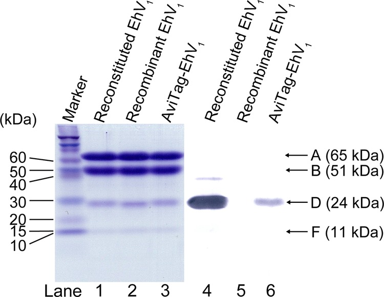
FIGURE 1.

Gel electrophoresis. Lanes 1–3, SDS-PAGE of reconstituted EhV1, recombinant EhV1, and AviTag-EhV1. A 16% gel was used; 12 pmol of protein was loaded in each lane. The molecular masses of the A, B, D, and F subunits are 65, 51, 24, and 11 kDa, respectively. Proteins were stained with Coomassie Brilliant Blue. Lanes 4–6, immunoblots stained by streptavidin-alkaline phosphatase conjugates, showing biotin labeling of the D subunit. Lane 4, reconstituted EhV1 containing the biotinylated D subunit. Lane 5, non-biotinylated recombinant EhV1. Lane 6, AviTag-EhV1 containing the biotinylated D subunit.
