
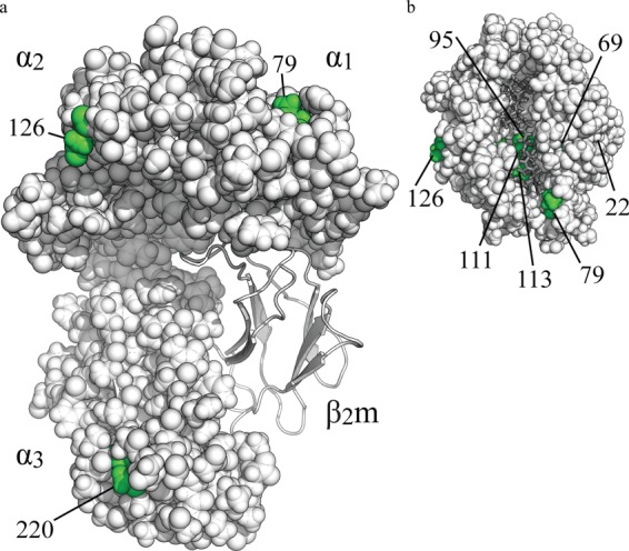
FIGURE 1.
Model of BF2*1501, depicting the location of the eight amino acids that differ between BF2*1501 and BF2*1901. The BF2*1501 structure is based upon the BF2*2101 structure (25) and is shown in a space-filling format with polymorphic amino acids shown in green. β2-Microglobulin (β2m) is shown in gray in a ribbon format, and peptide is shown in dark gray. a, side view; b, view from above the peptide binding groove. Polymorphic residue 22 is buried beneath the α1 helix. The side chains of residues 95 and 111 are on separate β strands with their side chains orientated toward each other.