Fig. 1.

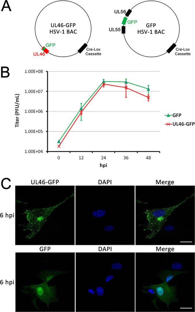
Characterization of a GFP-tagged pUL46 HSV-1 virus. A, Schematic of the BACs encoding for pUL46-GFP or free GFP-expressing HSV-1. B, pUL46-GFP HSV-1 grows with similar kinetics to free GFP-expressing HSV-1. Multistep growth curves were performed for both viruses in HFF cells. Cell-associated virus was harvested and titered at each time point by plaque assay. Error bars indicate S.E. from three biological replicates. C, pUL46-GFP accumulates to perinuclear and cytoplasmic punctae. Cells were infected with pUL46-GFP HSV-1 at an MOI of 0.5 and fixed at 6 hpi for direct fluorescence imaging. Scale bar demarcates 25 μm.
