
Fig. 2.
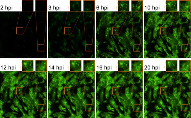
Live-cell imaging captures dynamic pUL46-GFP localization. HFFs were infected with pUL46-GFP HSV-1 at an MOI of 10 and imaged by epifluorescence time-lapse microscopy from 2 to 24 hpi. Magnified inserts indicate perinuclear accumulations.
Official websites use .gov
A
.gov website belongs to an official
government organization in the United States.
Secure .gov websites use HTTPS
A lock (
) or https:// means you've safely
connected to the .gov website. Share sensitive
information only on official, secure websites.
Live-cell imaging captures dynamic pUL46-GFP localization. HFFs were infected with pUL46-GFP HSV-1 at an MOI of 10 and imaged by epifluorescence time-lapse microscopy from 2 to 24 hpi. Magnified inserts indicate perinuclear accumulations.