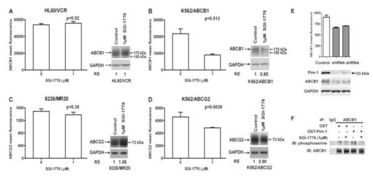
Figure 6. SGI-1776 treatment decreases surface expression of ABCB1 and ABCG2 on cells with high, but not low, Pim-1 expression.
A-D. HL60/VCR and K562/ABCB1 cells, expressing ABCB1, and 8226/MR20 and K562/ABCG2 cells, expressing ABCG2, were plated at a density of 1 × 10⁴ cells/ml with and without SGI-1776 1 μM for 48 hours, re-fed with fresh medium with and without the drug and cultured for 48 additional hours. ABCB1 surface expression was measured on 1 × 10⁵ HL60/VCR (A) and K562/ABCB1 (B) cells and ABCB1 surface expression on 1 × 10⁵ 8226/MR20 (C) and K562/ABCG2 (D) cells by flow cytometry, as detailed in Materials and Methods. Data are mean ± SEM of triplicate values per treatment. Protein lysates were prepared from the remaining cells and total ABCB1 or ABCG2 expression was quantified by densitometric analysis following immunoblotting. Immunoblots of ABCB1 or ABCG2 and GAPDH loading controls are shown. Relative expression (RE), calculated as detailed in Materials and Methods, is shown. E. Pim-1 knockdown was performed in K562/ABCB1 cells as described in Materials and Methods, resulting in decreased cell surface expression of ABCB1. Cell surface expression, measured by flow cytometry, is shown graphically in the upper panel, with the lower panel showing immunoblots measuring Pim-1, ABCB1 and GAPDH expression. F. ABCB1 and control IgG were immunoprecipitated from crude membrane extracts of High-Five insect cells overexpressing ABCB1 and serine phosphorylation of the immunoprecipitated ABCB1 by recombinant GST-tagged Pim-1 kinase in the presence and absence of 1 μM SGI-1776 was studied using an in vitro kinase assay as described in Materials and Methods. The resultant immunoblot probed with antibodies against phosphoserine and ABCB1 is shown. .

