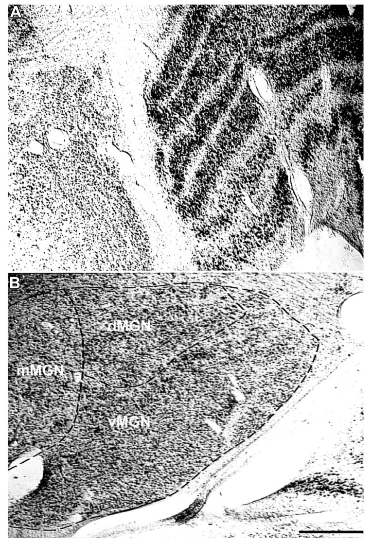
FIGURE 1.
Representative Nissl stains of the sensory thalamus. (A) Transverse Nissl stained section showing the anatomical proximity of the medial and lateral geniculate nuclei. (B) The three subdivisions of the MGN, the ventral (vMGN), dorsal (dMGN), and magnocellular (mMGN) MGN, defined according to previous conventions (Jones, 2003). All MGN sections were partitioned in this manner for stereological quantifications. The left sides of these images correspond to the medial aspect and the top to the dorsal aspect of the sections. Scale bar = 500 μm.

