Figure 3.

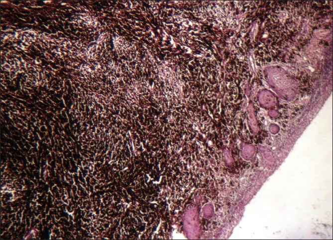
Photomicrograph of the lesion showing malignant melanocytes with extensive melanin pigementation infiltrating the connective tissue (H and E, ×10)

Photomicrograph of the lesion showing malignant melanocytes with extensive melanin pigementation infiltrating the connective tissue (H and E, ×10)