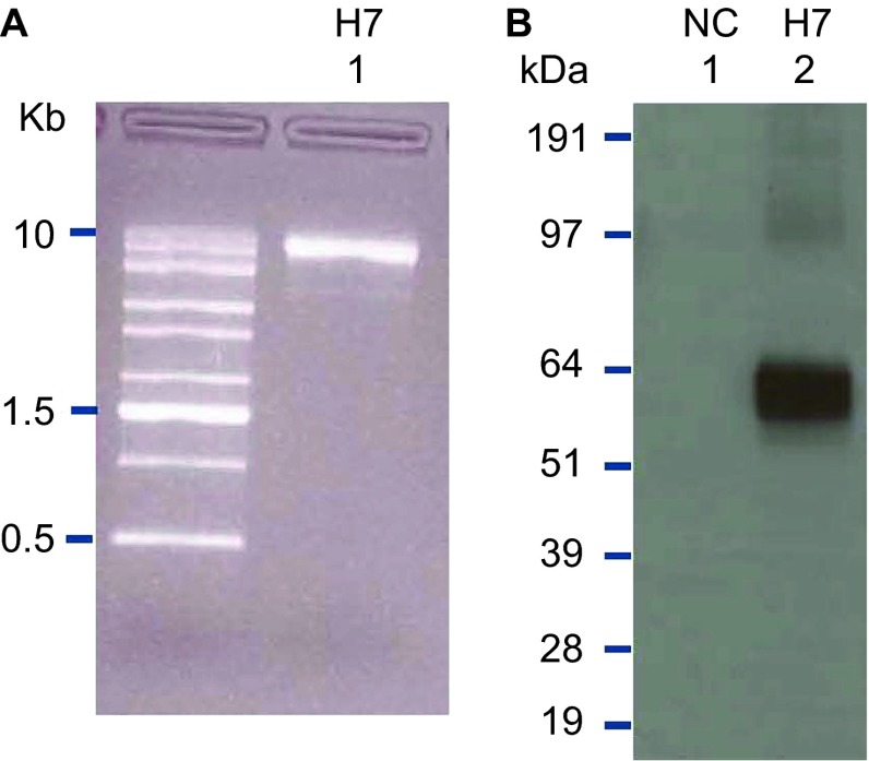
Figure 3.
RNA quality and confirmation of antigen expression. (A) The SAM vector encoding the H7 antigen was analyzed on a denaturing agarose gel. (B) A SAM vector encoding the H7 antigen was transiently transfected into BHK cells. Cell lysates were separated by SDS–PAGE, and blotted to nitrocellulose membranes. H7 HA was visualized by Western blot using a H7-specific polyclonal antibody. A SAM vector encoding eGFP served as an NC. eGFP, enhanced green fluorescent protein; NC, negative control; SDS–PAGE, sodium dodecyl sulfate–polyacrylamide gel electrophoresis.

