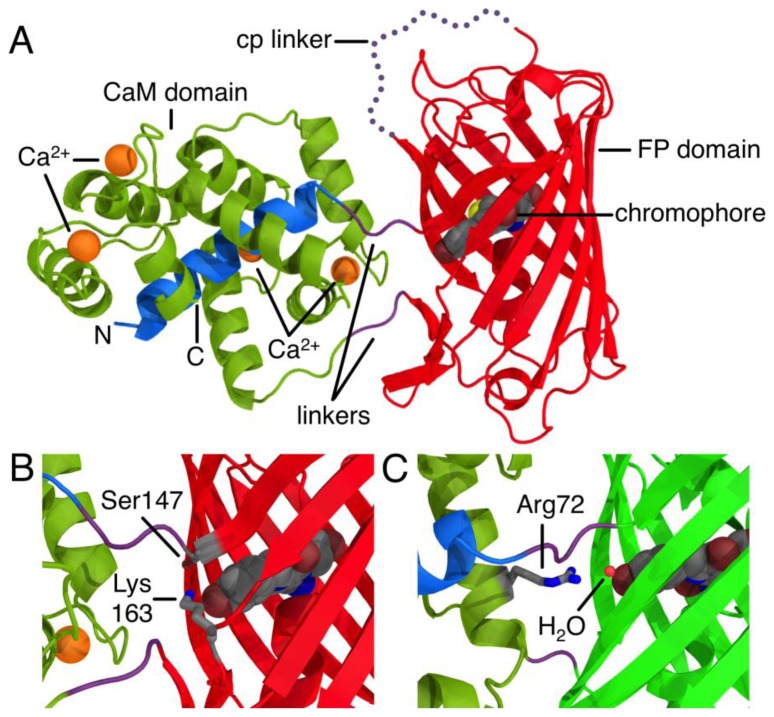
Figure 1.
The structure of a single FP-based Ca2+ indicator. (A) Cartoon representation of the X-ray crystal structure of R-GECO1 [8]. Domains and linkers are colored according to the sequence alignment shown in Figure 2. The “cp linker” is the linker used to connect the original N- and C-termini of the circularly permuted (cp) FP. (B) Zoom in on the circular permutation site of R-GECO1, where the chromophore is exposed to the interface between the CaM and FP domains. (C) The circular permutation site of GCaMP2 (PDB ID 3EVR) represented from a similar perspective to (B) [9].

