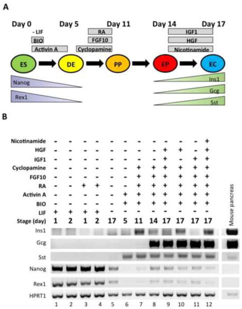
Figure 1. Differentiation of WT ES cells to pancreatic endocrine cells.
(A) Schematic representation of the endocrine differentiation protocol (14). WT murine ES cells are treated with different growth factors to differentiate the cells into definitive endoderm (DE), pancreatic progenitor (PP), endocrine progenitor (EP), and endocrine cells (EC). (B) WT ES cells were subjected to the 17-day differentiation protocol. Each lane represents a different condition at a specific time point. RT-PCR analyses were performed to monitor the expression of pancreatic differentiation markers such as insulin-1 (Ins1), glucagon (Gcg), somatostatin (Sst), neurogenin-3 (Ngn3), Pdx1, and Sox17, as well as the ES cell markers Nanog and Rex1. HPRT1 was used as a loading control. Pancreas extracts from C57BL/6 WT mice were used as a positive control (far right lane). Sample lanes are labeled 1–12 at the bottom. This experiment was performed 7 times, starting with fresh cells, with similar results.

