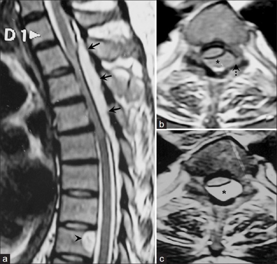
Figure 1.

a: T2-weighted, mid-sagittal magnetic resonance image revealing a thoracic, hyperintense, solitary, extradural mass (black arrows) opposite the D2-4 vertebral bodies. The signal intensity differs from that of the cerebrospinal fluid, but is similar to that of a remote intraosseous hemangioma within the D8 vertebral body. Axial magnetic resonance images reveal an eccentrically placed dorsal extradural mass (asterisk), which is isointense on T1- (b) and hyperintense on T2- (c) weighted images, with left foraminal extension (black dashed arrow)
