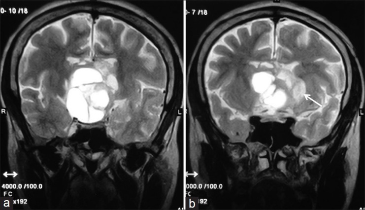
Figure 2.

Coronal T2-weighted MR images showing multicystic suprasellar mass lesion, with peripheral small solid component (arrow), which is showing hyperintense signal. The lesion is superiorly compressing the third ventricle extending upto the lateral ventricles. Optic chiasma is not seen separately from the lesion
