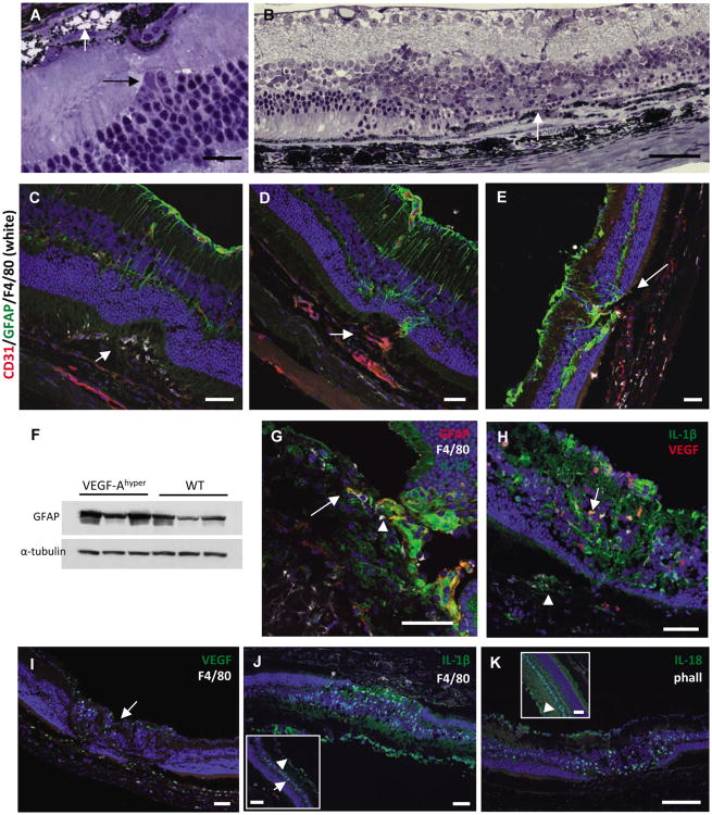
Figure 5. Glia cell activation in CNV lesions in VEGF-Ahypermice.
a. In young VEGF-Ahyper mice (6 weeks old) early RPE degeneration is seen only focally with someRPE cells undergoing vacuolar degeneration (intracellular vacuoles with remaining pigmentgranules; black arrow). At this stage retinal changes are limited, but migration of retinal Muller gliacells (white arrow) towards sites of RPE degeneration can already be observed. Scale bar 20μm.
b. In aged VEGF-Ahyper mice the photoreceptor layer adjacent to CNV lesions is present, butattenuated. Complete loss of photoreceptors is seen in retina overlying CNV lesions with anaccumulation of retinal Muller glia cells (arrow). Scale bar 50μm. 21 months old VEGF-Ahyper mouse.
c. At sites of early evolving CNV lesions subretinal macrophages (F4/80+, white; arrow) are seenprior to neovessel formation. Limited to the overlying retina GFAP expression (green) increases,indicating glia cell activation and proliferation, while adjacent retina appears unchanged. Scale bar50μm. 4 months old VEGF-Ahyper mouse.
d. In early CNV lesions with subretinal neovascularization (CD31, red; arrow) GFAP+ glia cells have increased and have infiltrated early CNV lesions (green). Scale bar 50μm. 4 months old VEGF-Ahyper mouse.
e. In aged VEGF-Ahyper mice with large CNV lesions, massive GFAP expression and glia cell infiltration into sites of CNV lesion formation is seen (arrow). Scale bar 50μm. 15 months old VEGF-Ahyper mouse.
f. Retinal GFAP is increased in aged VEGF-Ahyper mice compared to littermate control mice. 16 months old mice.
g. Co-immunolabeling for GFAP (red), IL-1β (green) and F4/80 (white), shows that high IL-1β expression is seen in infiltrating retinal glia cells (arrowhead), while lower levels are observed in RPE cells and only in few macrophages in CNV lesions (arrow). No IL-1β is detected in most choroidal macrophages. Scale bar 50μm. 15 months old VEGF-Ahyper mouse.
h. Retinal glia cells co-express both VEGF-A and IL-1β in the retina overlying CNV lesions where the photoreceptor layer is severely attenuated (arrow). Labeling for IL-1β and VEGF-A is also seen in RPE cells (arrowhead) in CNV lesions. Scale bar 50μm. 15 months old VEGF-Ahyper mouse.
i. Increased VEGF-A expression (green) is seen in retinal glia cells overlying CNV lesions (arrow). Scale bar 50μm. 15 months old VEGF-Ahyper mouse.
j. While IL-1β is expressed in glia cells overlying CNV lesions that have replaced the ONL, adjacent normal appearing retina (inset) shows only weak IL-1β expression in the INL (arrow) and GCL (arrowhead). Scale bars 50μm. 15 months old VEGF-Ahyper mouse.
k. Infiltrating retinal glia cells show strong expression of IL-18 overlying CNV lesions. Scale bar 50μm. 15 months old VEGF-Ahyper mouse. See also Figure S5.

