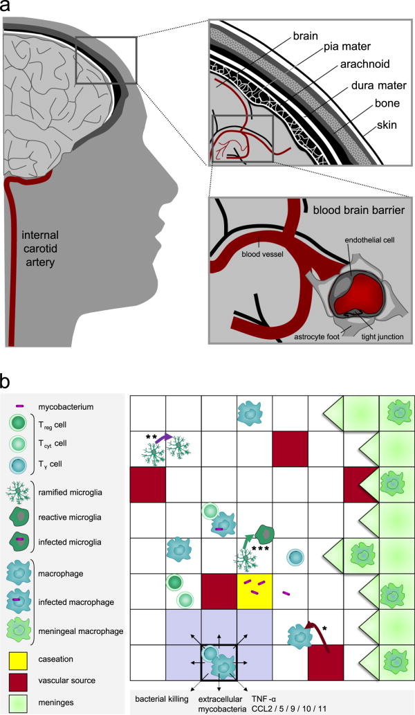
Figure 2.
Brain environment. (a) Environment of TBM. The right upper panel shows a cross-section of brain, meninges, skull and skin. The right lower panel shows cerebral blood vessels with a detail of the blood-brain barrier: the blood vessel wall consists of closely-connected endothelial cells resulting in tight junctions that are supported by astrocytes. (b) Environment of the granuloma showing a part of the 10,000 micro-compartments grid. In the panel different immune cells, bacteria and static properties of the grid are depicted. Macrophages and T cells enter the grid through vascular sources (*). Microglia proliferate on site (**). Example of microglia state transition from ramified to reactive (***). The Moore neighborhood of the micro-compartment with highlighted borders is colored in gray.

