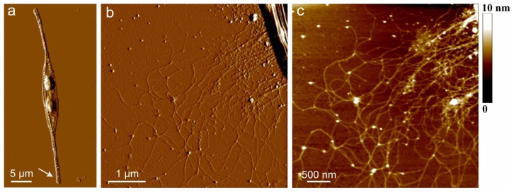
Figure 1.
Extracellular polymers released by C. closterium (CCNA1) obtained by AFM imaging in contact mode after deposition on mica surface. (a) AFM image of the whole cell presented as deflection data. The arrow indicates the position of the polymer excretion site; (b) The released polymers still attached to the apex of the cell rostrum, deflection data, scan size 5 μm × 5 μm; (c) Released polymers presented as height data, scan size 4 μm × 4 μm and vertical scale shown as the color bar (reproduced from [33]).

