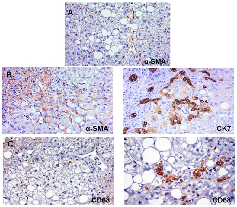
Figure 1.
(A) Immunohistochemistry for α-smooth muscle actin (α-SMA) in non-alcoholic fatty liver disease (NAFLD). In NAFLD, pericentral fibrogenesis is due to activation of hepatic stellate cells (HSCs) which acquire α-SMA positivity. Original Magnification: 20×; (B) Immunohistochemistry for α-smooth muscle actin (α-SMA) and Cytokeratin (CK)-7 in NAFLD. In advanced stages of NAFLD, periportal fibrogenesis is present. In this case, α-SMA positive myofibroblasts surround CK7+ reactive ductules at the periphery of portal spaces. Original Magnification: 20×; (C) Immunohistochemistry for CD68 in NAFLD. CD68 is specifically expressed by Kupffer cells and macrophages which are distributed throughout entire liver lobule both at pericentral and periportal position. Macrophage foam cells are clearly recognized in left image. Original Magnification: 20× (right) and 40× (left).

