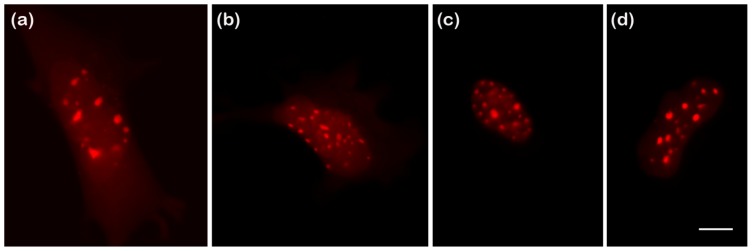
Figure 4.
Epifluorescent images of GHFT1 cells expressing FP-BZip fusion proteins. All four red FP-BZip fusion proteins had the expected localization to regions of heterochromatin. FCS data for Figure 3c,d were obtained in the cytosol and in the nucleus away from the heterochromatin bundles. (a) mCherry-BZip; (b) mApple-BZip; (c) TagRFP-T-BZip; and (d) mRuby2-BZip. Scale bar = 10 μm.

