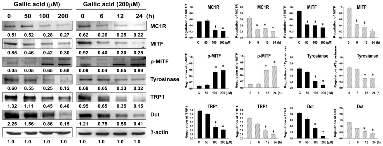
Figure 3.
Expressions of melanogenesis-related proteins in B16F10 cells with gallic acid treatment. Western blotting data show the changes in MC1R, MITF, p-MITF, tyrosinase, TRP1, and Dct expressions in B16F10 melanoma cells treated with gallic acid at different concentrations (0–200 μM) for 24 h and treated with 200 μM of gallic acid at different times. β-Actin was used as the protein loading control. Statistical results represented as Means ± SEM (n = 3) by ANOVA with the Tukey-Kramer test (* p < 0.001 compared with the control).

