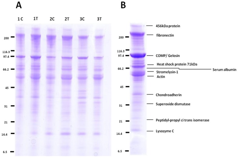
Figure 1.
1D-SDS-PAGE of the cartilage OA secretomes demonstrated little difference in the profiles following IL-1β treatment. (A) OA human articular cartilage explants (n = 3) were cultured in media supplemented with 10 ng/mL IL-1β (T) or un-supplemented media (C). Culture media were collected at two days for further analysis by SDS-PAGE and staining with Coomassie Brilliant Blue. Equal protein loading of 20 μg of protein per well allowed a qualitative comparison of the secretomes; (B) the most abundant proteins in the media marked at the positions of the bands were excised from the gel, trypsin digested, and the protein content of each single band was analysed using peptides identified using LC-MS/MS. Proteins indicated on the gel correlate to the size and are the primary protein identified in the corresponding gel analysis.

