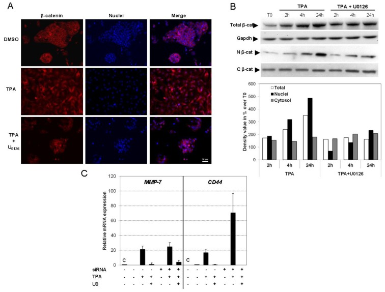
Figure 7.
TPA promotes EMT through concomitant activation of the Snaill and Wnt/β-catenin pathways. (A) HepG2 cells were grown on coverslips and treated with TPA with or without U0126 for 48 h. After exposure, the cells were fixed and processed for indirect immunofluorescence analysis for the detection of β-catenin (red) and visualization of nuclei (DAPI, blue); (B) At indicated time, nuclear (N) and cytosolic (C) fractions were prepared as described in materials and methods, and β-catenin was detected by Western blotting. The semi-quantification of chemiluminescence was performed after the acquisition with a CCD camera. Results are expressed as a percentage of T0-treated cells, designated as 100%. The results are representative of three independent repeats for each experiment; (C) Changes in mRNA levels for MMP-7 and CD44 genes upon TPA treatment were assessed by real-time RT-PCR after transfection of control siRNA (C) or Snail siRNA. The HepG2 cells were stimulated with 100 nM TPA, with or without U0126 for 48 h. Relative mRNA expression levels (normalized with respect to gapdh) were determined and mRNA levels in DMSO-treated cells (untransfected condition) were set to 1. Error bars indicate the means ± SEM of triplicate determinations from three independent experiments.

