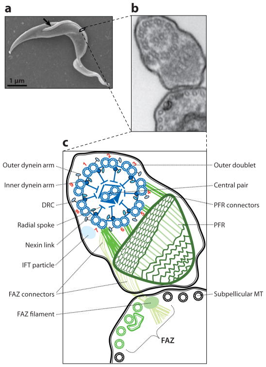
Figure 1.
The trypanosome flagellum. (a) Scanning electron micrograph (EM) of a procyclic trypanosome. The arrow indicates the single flagellum. (b) Transmission EM showing a cross-section of the flagellum and its attachment to the cell body as viewed looking from posterior toward anterior. (c) Schematic representation of the micrograph in panel b. Major flagellar substructures are indicated and outer doublet microtubules are numbered according to convention. Flagellar substructures that are broadly conserved among eukaryotes are in blue, and structures that are unique to trypanosomes and closely related organisms are in green. Abbreviations: DRC, dynein regulatory complex; FAZ, flagellum attachment zone; IFT, intraflagellar transport; MT, microtubule; PFR, paraflagellar rod. Adapted from Reference 113, with permission.

