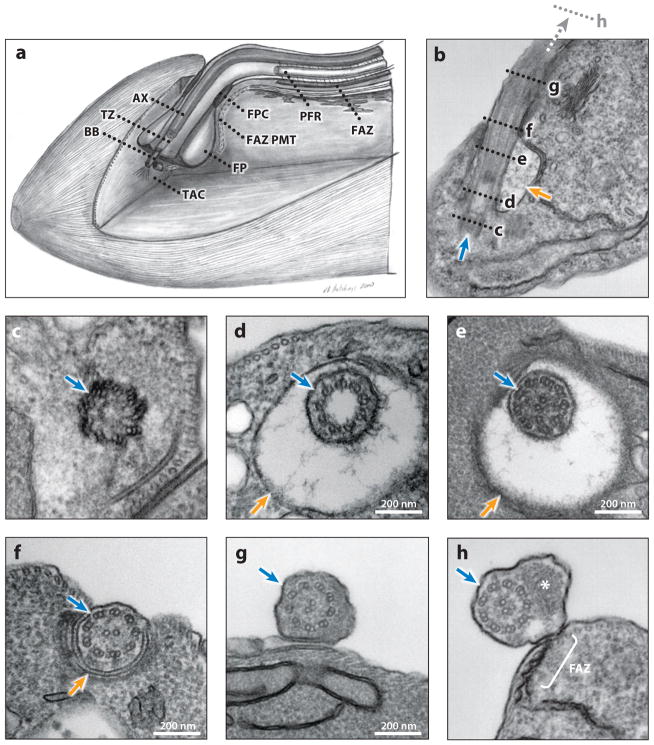
Figure 2.
Ultrastructure of the trypanosome flagellum. (a) Schematic cut-out view of the flagellar pocket area. Note that because the mitochondrion is not shown, only a partial TAC, consisting of the exclusion zone filaments extending from the basal body toward the mitochondrion, is represented in this schematic. (b–h) Electron micrographs of longitudinal (b) and transverse (c–h) sections of procyclic cells, from posterior (c) to anterior (h). (b) Longitudinal section showing the flagellar apparatus (blue arrow) and FP (orange arrow). The positions of the transverse sections in panels c–h are indicated with dashed lines, with panel h farther anterior than panel g. (c) The basal body apparatus. (d) The 9 + 0 transition zone within the FP; note the filaments of the ciliary necklace radiating from the doublet microtubules. (e) The 9 + 2 flagellar axoneme within the FP. (f) The flagellum exiting the FP through the FPC. (g) The flagellum outside of the FP but not yet having a PFR. (h) The flagellum outside of the FP, containing both an axoneme and a PFR (asterisk). Note that FAZ connections to the axoneme initiate proximal to the initiation of the PFR (not shown). Panel a is adapted from Reference 55, with permission. Abbreviations: AX, 9 + 2 axoneme; BB, basal body; FAZ, flagellum attachment zone; FAZ PMT, the four specialized subpellicular microtubules of the flagellum attachment zone; FP, flagellar pocket; FPC, flagellar pocket collar; PFR, paraflagellar rod; TAC, tripartite attachment complex; TZ, transition zone.

