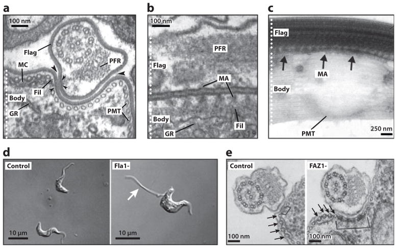
Figure 3.
The flagellum attachment zone (FAZ). (a) Transverse electron micrograph (EM) section of the Trypanosoma brucei rhodesiense bloodstream form, showing the flagellum (Flag) and cell body (Body). The electron-dense filament (Fil), macula adherens (small arrowheads) and membranous compartment (MC) of the FAZ are indicated. Large arrowheads point out the gap between the flagellar and cell body membranes. Also indicated are the subpellicular microtubules (PMT), the paraflagellar rod (PFR), and the granular endoplasmic reticulum (GR). (b) Longitudinal section of T. vivax, showing a series of macula adherens (MA) with their anchoring strands passing to longitudinally oriented filaments. (c) Whole mount of an intact, detergent-extracted T. brucei cytoskeleton, with the MA indicated with arrows. (d) RNAi knockdown of Fla1 in bloodstream-form T. brucei cells (Fla1-) leads to flagellar detachment (arrow). (e) RNAi knockdown of FAZ1 in procyclic T. brucei cells (FAZ1-) leads to abnormalities in FAZ ultrastructure, including unusually wide FAZ filaments (bracket). Arrows point to the four specialized PMTs of the FAZ. Panels a and b are adapted from Reference 145, panel c from Reference 58, panel d from Reference 69, and panel e from Reference 142, with permission.

