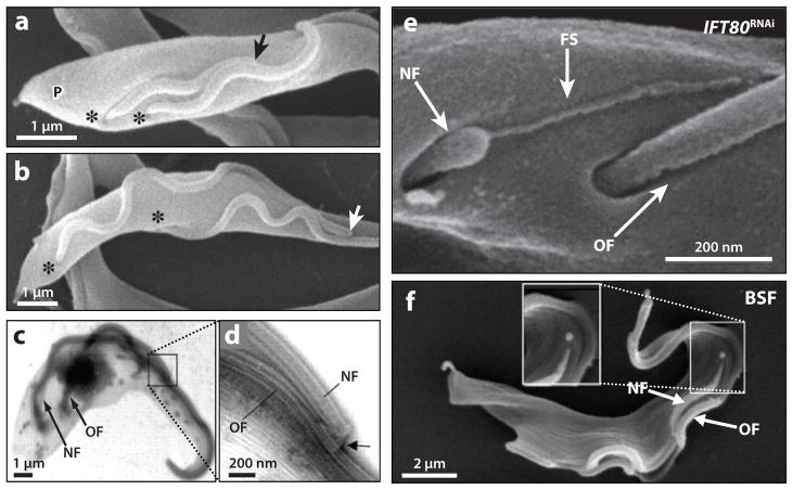
Figure 4.
Trypanosome flagellum biogenesis. (a,b) Scanning electron micrographs of procyclic-form cells show that the distal tip of the new flagellum (arrow) remains associated with the old flagellum during cell division. The distal tip of the new flagellum is indicated (arrow), the flagellar pockets are marked (asterisks), and the cell posterior is indicated (P). (c,d) Low (c) and high (d) magnification transmission electron micrographs of a detergent-extracted procyclic cytoskeleton showing the flagellar connector (arrow) that is present at the tip of the new flagellum (NF) and contacts the old flagellum (OF). (e) Scanning electron micrograph of a procyclic-form IFT80 RNAi knockdown mutant. A flagellar membrane sleeve (FS) of the new flagellum (NF) extends toward the lateral aspect of the old flagellum (OF). (f) Scanning electron micrograph of a bloodstream-form (BSF) cell during cell division, demonstrating that the tip of the new flagellum (NF) does not appear to connect to the old flagellum (OF) during elongation. Panels a–d are adapted from Reference 83, panel e from Reference 25, and panel f from Reference 16, with permission.

