Figure 5.
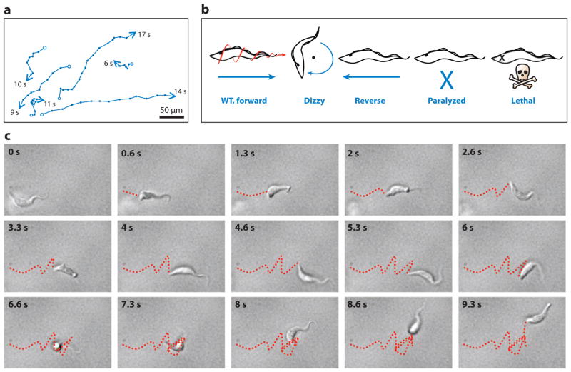
Trypanosoma brucei cell motility. (a) Motility traces of wild-type procyclic cells. The positions of individual cells are plotted at 1-s intervals (indicated by dots). Starting positions are marked with open circles; ending positions are marked with arrowheads, and the time in seconds that each cell was within the field of view is indicated. (b) Schematic representations of motility phenotypes in procyclic motility mutants. Wild-type (WT) cell movement is helical with the flagellum tip leading. Dizzy mutants, such as trypanin knockdown mutants (58), retain a vigorously beating flagellum but are unable to move directionally. Reverse mutants, such as DNAI1 (15) or LC1 (9) knockdown mutants, exhibit a reverse, base-to-tip beat, and move backward, with the flagellum tip trailing. Paralyzed mutants, such as central pair and radial spoke mutants (15, 114), are incapable of movement at the cellular level. Lethal defects arise in mutants with severe flagellar paralysis (10). (c) Time series (0.66-s intervals) of a WT procyclic cell moving in liquid medium. The movements of the posterior tip of the cell body are traced with a red dashed line. Note that periods of running (0–4.66 and 8–9.33 s) alternate with periods of tumbling (5.33–7.33 s). Panel a is adapted from Reference 58, and panel b from Reference 113, with permission.

