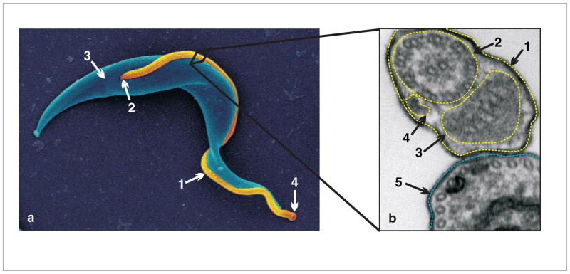
Fig. 1. Trypanosoma brucei cell and flagellum structure.

(a) Scanning electron microscopy image of PCF T. brucei. Cell body pseudocolored in blue and flagellum in gold.1 flagellum, 2 flagellar pocket, 3 cell body posterior end, 4 flagellar tip, corresponds to cell anterior. (b) Cross-section of PCF T. brucei cell as imaged by transmission electron microscopy. 1 flagellar membrane, 2 axoneme, 3 paraflagellar rod, 4 Intraflagellar transport particle, 5 cell body membrane. Subpellicular microtubules (not labelled) are visible in cross section in cell body. Adapted from Ralston et al. 2009. Ann. Review. Microbiol, with permission.
