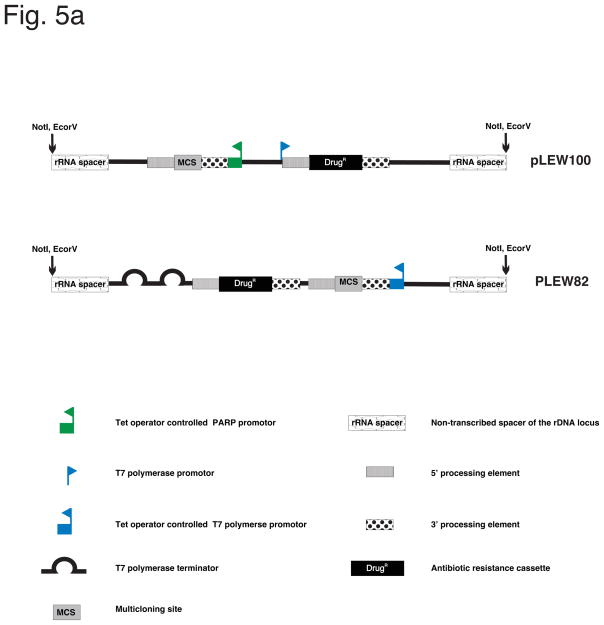
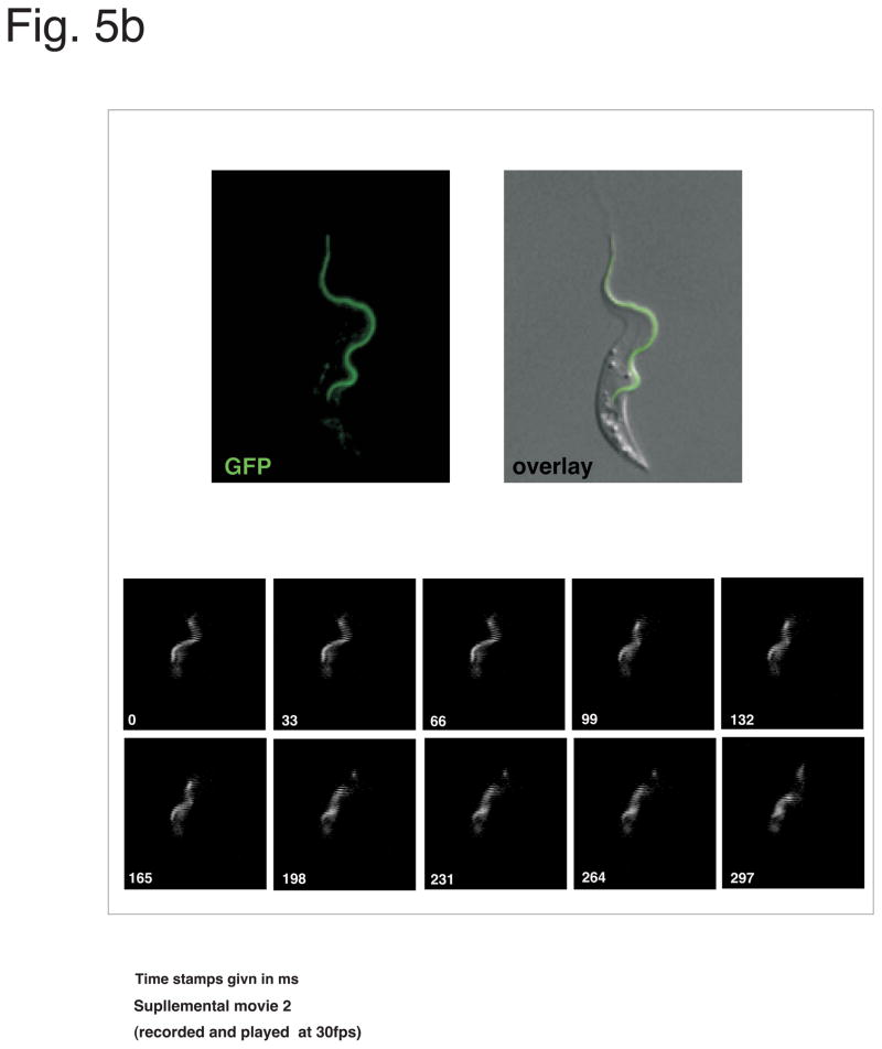
Fig. 5. Plasmids for inducible gene expression.
(a) Illustration of two vectors (pLEW100 and pLEW82), commonly used for inducible expression in T. brucei. Adapted from Wirtz et al. 1999, MBP, with permission. (b) Example of a PCF cell line expressing a GFP-tagged flagellum protein (PFR2) through the use of pLEW100 (Fig. 5a). Top panels, fluorescence image of a Tet induced, live cell. On bottom, series of images showing the movement of the GFP-PFR2 expressing cell line under fluorescence microscopy (see supplementary movie 2. Cells are co-stained with a live DNA dye, CFSE to visualize nucleus and kinetoplast. Movie is recorded using a 63× objective (recorded and played back at 30fps).


