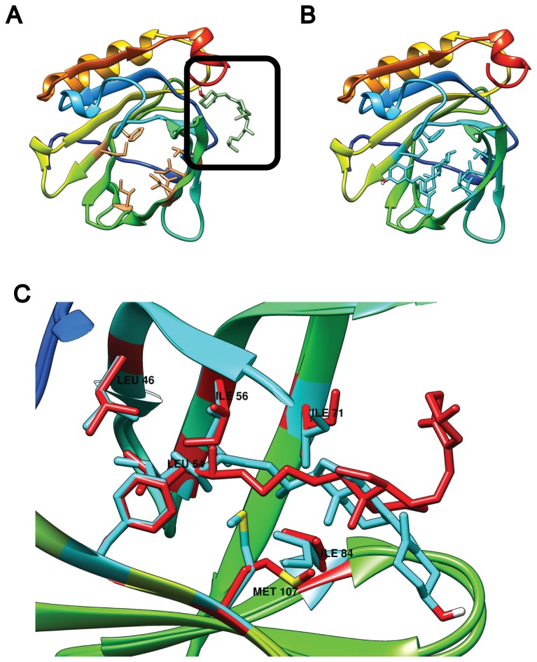
Figure 3. Effect of residue flexibility on VD3 binding to monomeric BLG.
(A) Docking to 2BLG with a rigid calyx locates VD3 to Site C (black square). In contrast, when seven residues are rendered flexible, VD3 fits in the calyx (B and C). In (C) a side view magnification of the calyx compares the VD3 docking result (blue) and the residues made flexible (blue), to their XRD counterparts (red).

