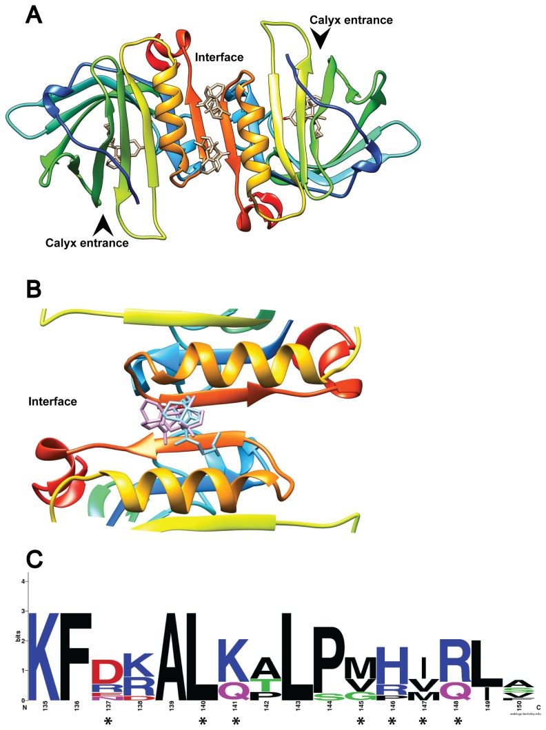
Figure 4. VD3 binding to the BLG dimer interface.
(A) The BLG dimer is shown with the four experimentally determined VD3 ligands at their respective binding sites. The arrowheads indicate entrance to the calyx. In (B) only one experimental VD3 (purple) is compared to the best docking result obtained with flexible residues (“full interface” in 2BLG, in blue). (C) Shows the weblogo of 7 ungulate BLG sequences highlighting the residues that bind VD3 with asterisks.

