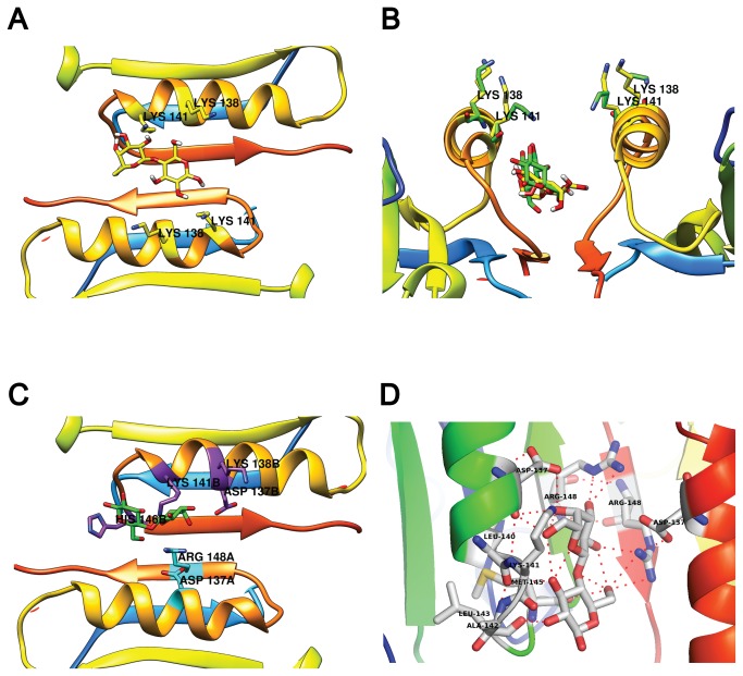
Figure 5. Lactose docking to the 2BLG dimer.
(A) Lactose docking to a rigid BLG dimer. Notice both K138 and K141 pointing away from lactose. In (B), a side view of the rigid interface docking (lactose, K138 and K141 in yellow) is compared to the “fully flexible” results (green) where K141 shifts towards lactose. In (C) a top view of the best “fully flexible” result. Residues involved in lactose binding are highlighted by chain: chain B in purple and chain A in cyan. (D) close-up of the interfacial binding site showing chains from both BLG monomers and their respective hydrogen bonds to lactose.

