
Fig. 7.
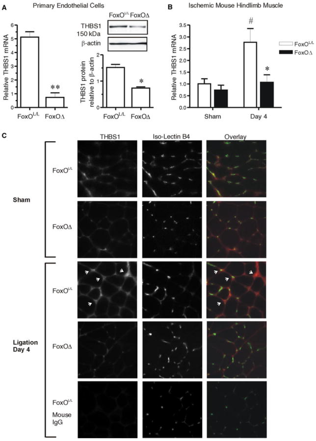
FoxO deletion prevents the ischemia-induced increase in Thrombospondin 1 within the endothelium of mouse muscle. Microvascular endothelial cells were isolated from skeletal muscle of FoxOL/L and FoxOΔ mice. THBS1 mRNA was assessed by q-PCR (normalized to Hprt1) and THBS1 protein a was assessed by Western blot (normalized to β-actin). **p = 0.004 compared to cells from FoxOL/L mice; *p = 0.01, paired Student’s t test, n = 3 independent isolations, from a total of 8 individual mice per condition. THBS1 mRNA gastrocnemius muscles from FoxOL/L and FoxOΔ mice b was assessed by q-PCR (normalized to Hprt1) at 4 days post-femoral artery ligation or corresponding sham operation. Two-way ANOVA shows main effects of FoxO deletion and arterial ligation (p = 0.04 and p = 0.03, respectively). Significant differences are #p < 0.05 versus Sham FoxOL/L, and *p < 0.05 versus day 4 ligated FoxOL/L (Bonferonni Post hoc, n = 4–7 mice). TA/EDL muscle cross-sections from 4 day ligated or sham operated FoxOL/L or FoxOΔ mice were immunostained c for THBS1 (red) and counterstained with Isolectin B4 (green) to identify capillaries. Arrows point to peri-capillary THBS1 immunoreactivity in the ischemic FoxOL/L tissue. Normal mouse IgG was used as a negative control for the THBS1 antibody